Promoting the Science of Tissue Regeneration |
Featured Images
Chen-Hui Chen Lab
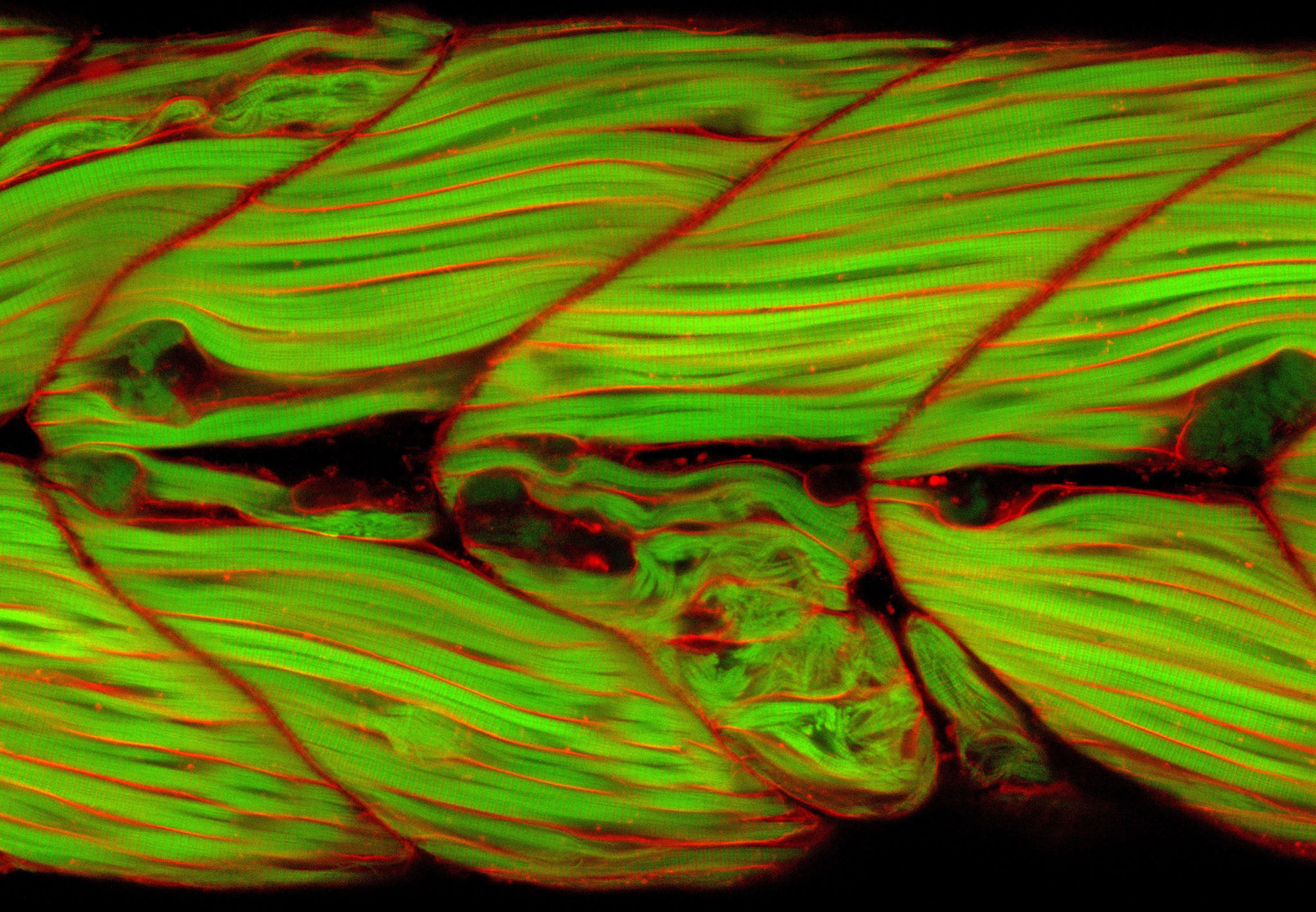
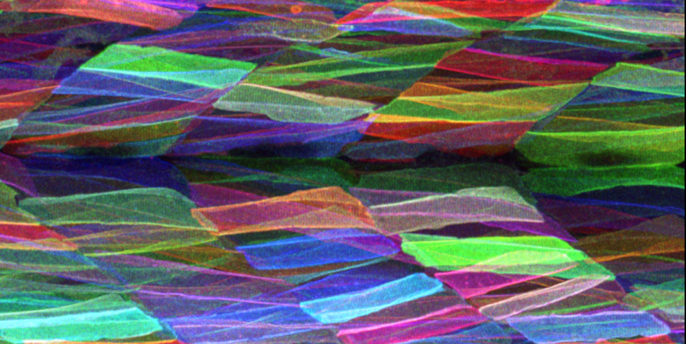
This image celebrates the colours of the rainbow with work from Uday Kumar, a PhD student in Chen-Hui Chen's lab at Academia Sinica in Taiwan. The Chen lab uses imaging and genetics to discover underlying regenerative principles. Here, Uday is studying muscle repair using a zebrafish model. Zebrafish myofibers are shown labelled using a brainbow technique where each myofiber is marked by a distinct color.(Image credit: Uday Kumar) Andrea Wills Lab
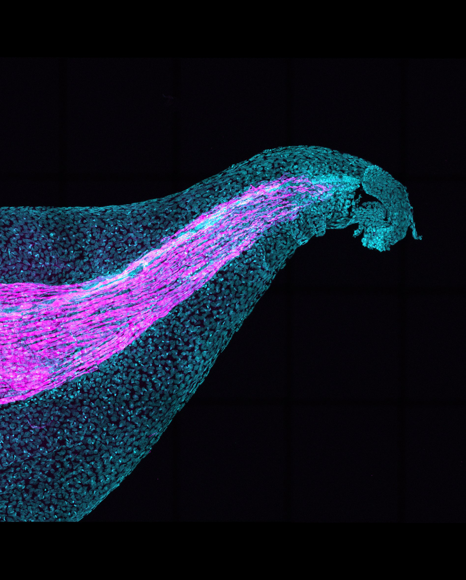
Tadpole tail tales. A major goal of the Wills lab (University of Washington) is to understand complex repair responses during tail regeneration in frog tadpoles. PhD student Jeet Patel studies how injury response directs tissue structure regeneration. Five days after truncation, the major architecture of the Xenopus tropicalis tail is restored. However, regenerated muscle fibers (pink) extend caudally instead of forming chevrons as in development. (Image credit: Jeet Patel) Elly Tanaka Lab
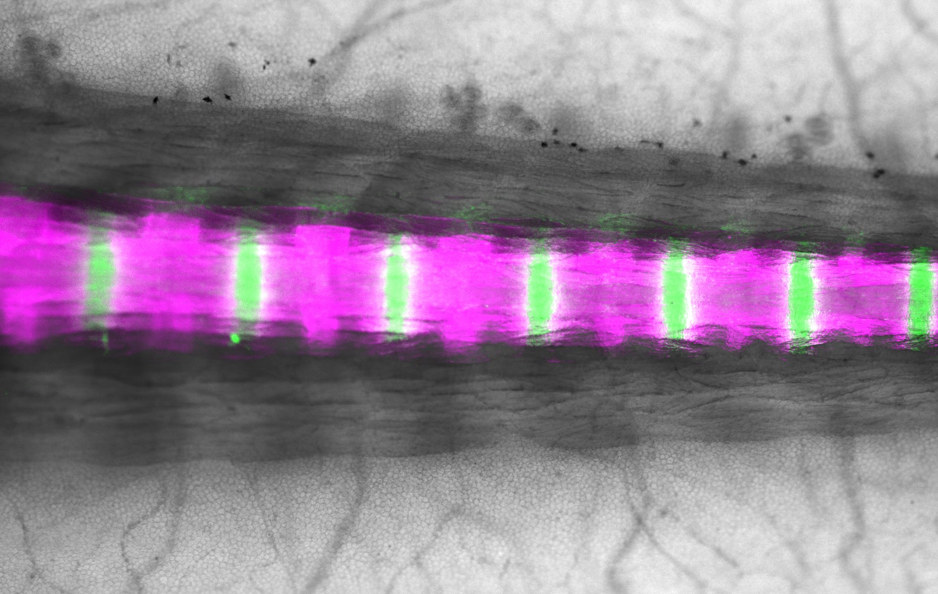
Vertebral column repair. Salamanders have the remarkable capacity to regenerate after injury. Wouter Masselink PhD, a postdoc in the Tanaka Lab at the IMP Research Institute of Molecular Pathology at the Vienna Biocenter (VBC) is interested in the underlying mechanisms. These are images of the uninjured and injured vertebral column illuminated by Collagen (Green) and Alizarin Red (Magenta). (Image credit: Gisela Deneke and Wouter Masselink) Didier Stainier Lab
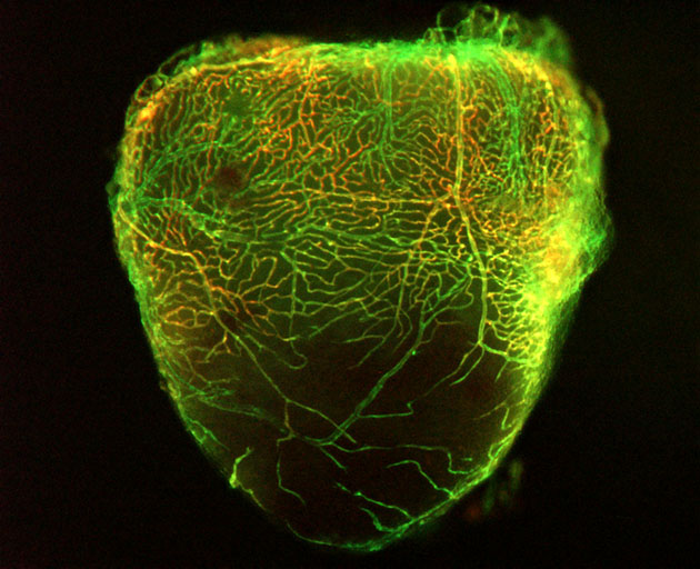
Mending injured hearts. A major goal of the Stainier lab at the Max Planck Institute for Heart and Lung Research is to understand mechanisms regulating cardiac regeneration. PhD student Hadil El-Sammak is studying revascularization of the zebrafish heart after cryoinjury. Here coronary endothelial cells (red and green) can be seen invading the injured area (bottom tip) during cardiac regeneration. (Image credit: Hamil El-Sammak) Sandoval-Guzmán lab
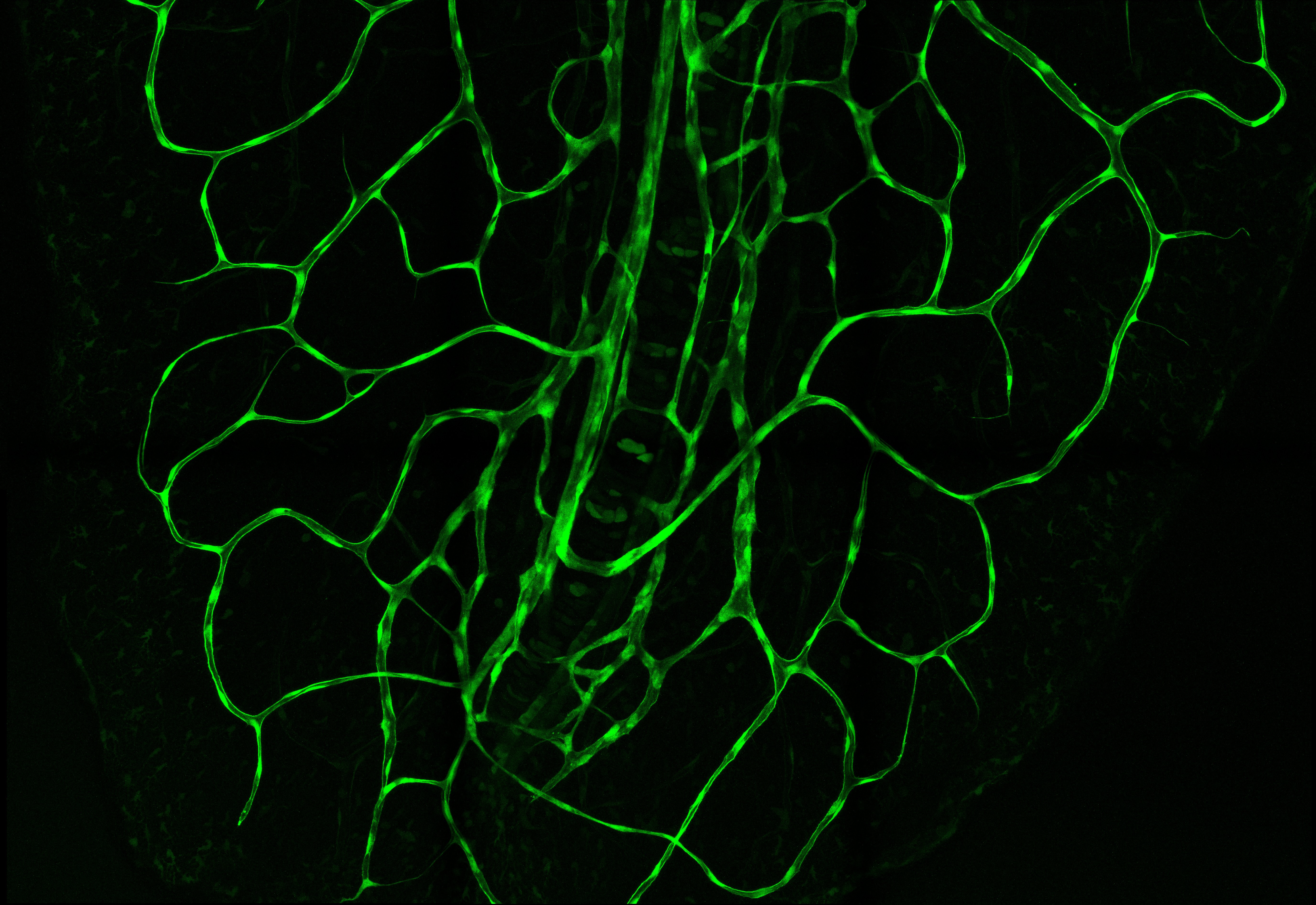
New vessels for regeneration. Camilo Riquelme Guzmán is a PhD Student in the Sandoval-Guzmán lab at Technische Universität, Dresden where he studies revascularization during axolotl limb regeneration. In this image, blood vessels in the axolotl (in this case the tail) can be visualized with GFP. Camilo Riquelme Guzmán is using this Tie2 transgenic line to study blood vessels during limb regeneration. | Alejandro Sánchez Alvarado Lab
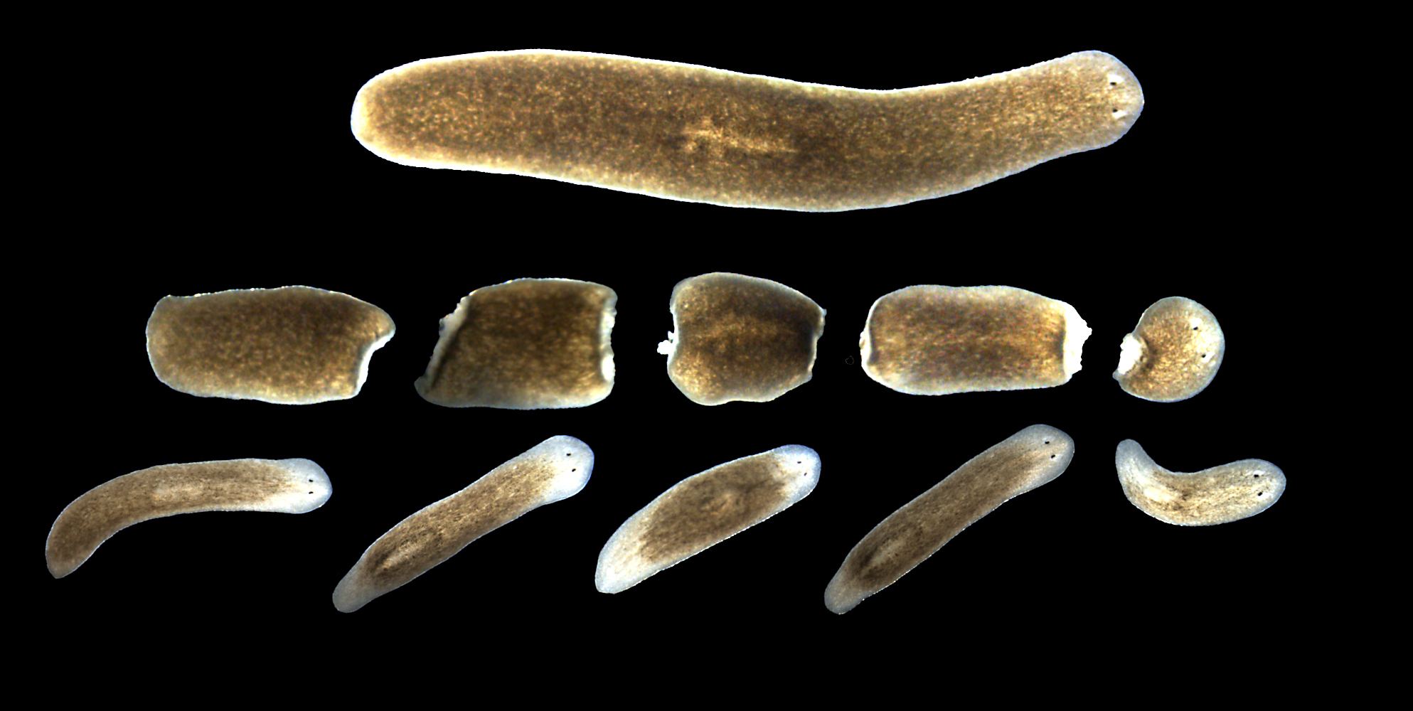
Research at the cutting edge. The planarian flatworm Schmidtea Mediterranea can regenerate a whole organism from just about any fragment of its body. This is the topic of research for Steph Nowotarski, PhD, a postdoc in the Sánchez Alvarado Lab at the Stowers Institute for Medical Research. This image shows the same animal intact, just cut, and two weeks later. (Image credit: Steph Nowotarski) Mimi Sammarco Lab
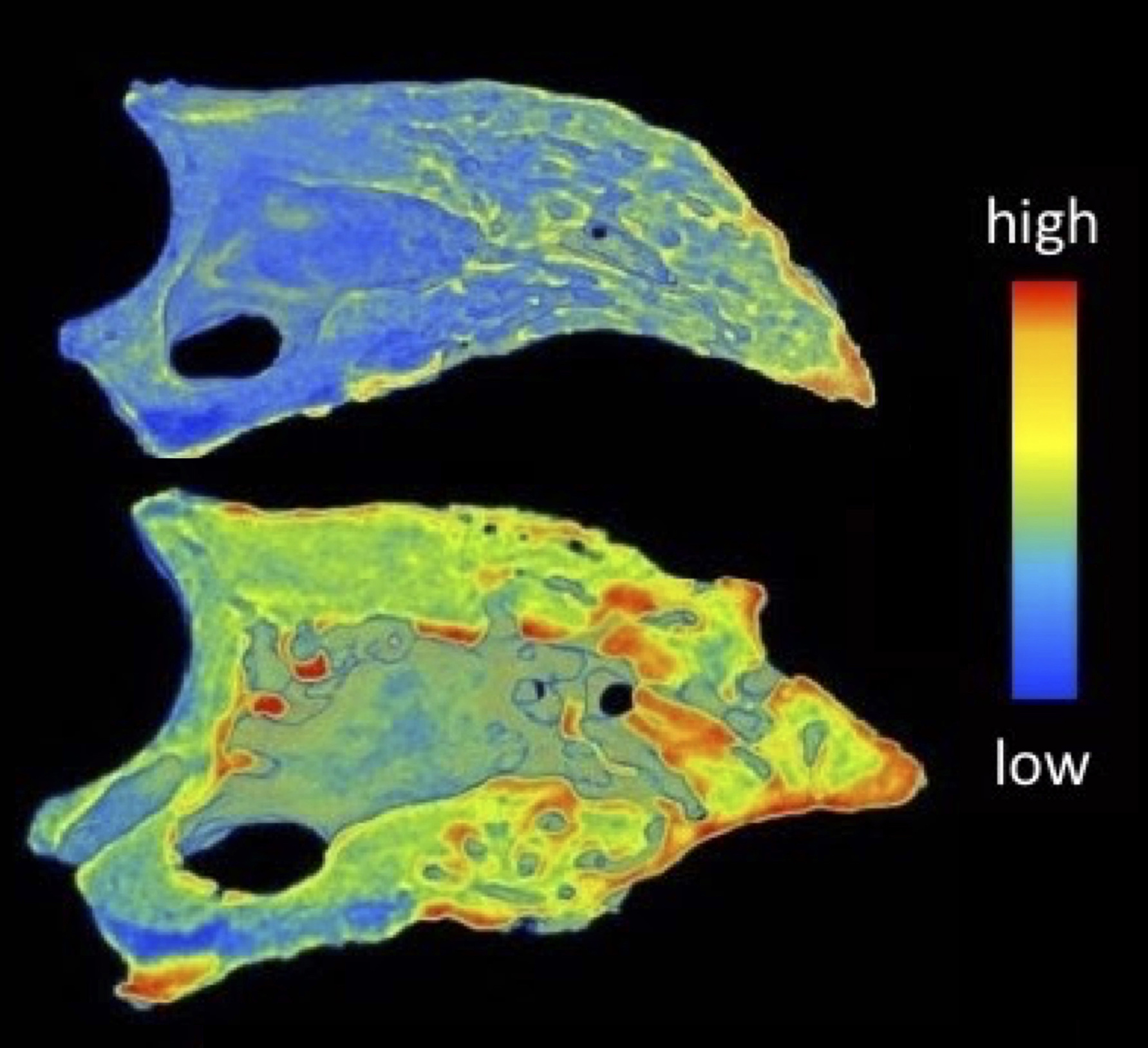
Visualizing bone regeneration at the digit tip. The Sammarco Lab at Tulane School of Medicine is developing high resolution micro-CT methods to assess bone mineral density during regeneration. Depicted are mouse digit tip bones that have undergone regeneration after amputation (42 days). The top depicts regeneration in a 6 month old mouse, while the bottom depicts regeneration in an 18 month old mouse. This imaging method allows for precise quantification of mineral density during bone regeneration. (Image credit: Mimi Sammarco, PhD) Sánchez Alvarado Lab
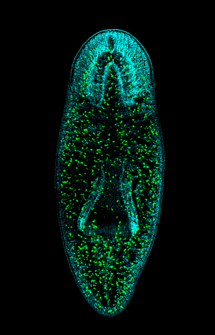
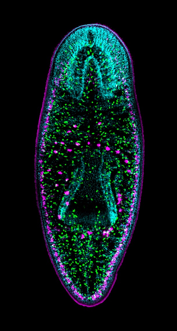
Constellations of stem cells in the regenerating planarian. Frederick Mann, PhD (Postdoc) and Julianna Haug (Research Associate) at the Stowers Institute in the Sánchez Alvarado Lab, are studying how cell fates change during whole body regeneration. These images depict single confocal sections of the asexual planarian Schmidtea mediterranea. Double fluorescent RNA in situ hybridization is used to mark subepidermal gland cells (reticulocalbin-2 in magenta) and epithelial precursor cells (prog-1 in green); DNA is stained with DAPI (cyan). (Image credit: Julianna Haug, Chris Arnold, PhD, and Carlos Guerrero-Hernandez.) Peter Currie Lab
Zebrafish muscles to the rescue. Jaochim Berger, PhD, a postdoc in the Currie Lab at the Australian Regenerative Medicine Institute, Monash University uses transgenic zebrafish to mark myofibrils (green) and the sarcolemma (red). Dystrophic muscle can easily be seen in a model for Duchenne’s Muscular Dystrophy providing a new way screen for therapeutics. (Image credit: Joachim Berger) Yun Lab
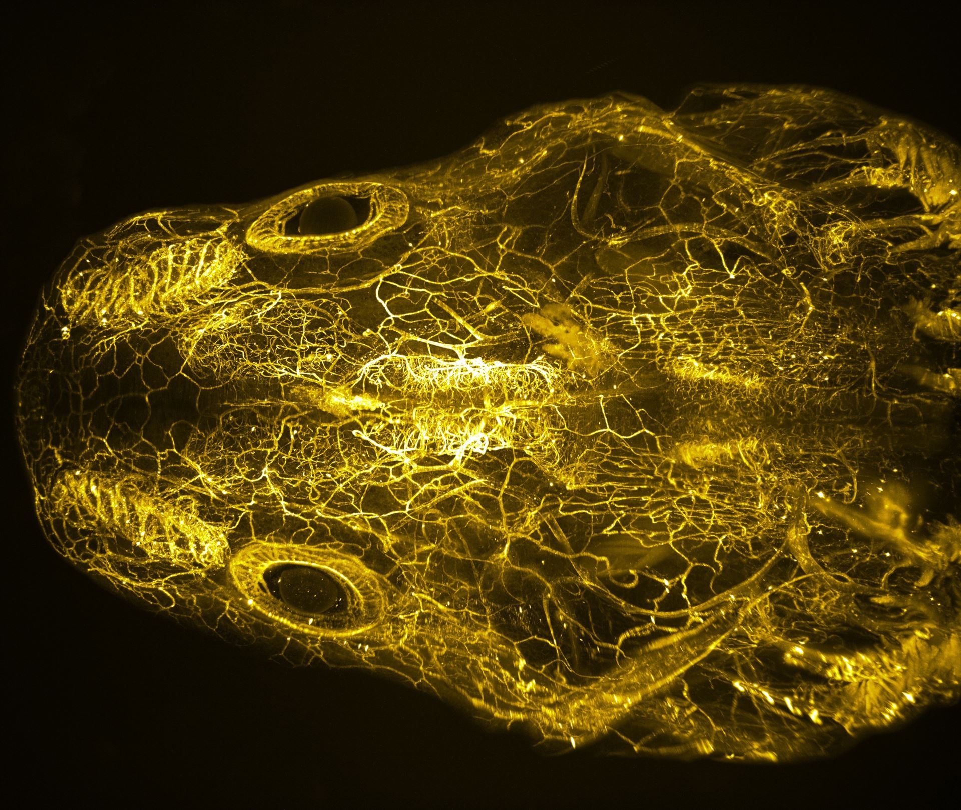
3D image of the vasculature of the axolotl head Lizbeth Airais Bolaños Castro is a PhD student in the Yun Lab at Technische Universität Dresden. She generated this 3D image of the vasculature of the axolotl head. The vasculature is mapped using lectins, following tissue clearing and light-sheet microscopy. Lizbeth is using this approach to characterise the vascularization during organ regeneration.
|
 Somewhere over the rainbow.